Visual evoked potential (VEP) and Electroretinogram (ERG) tests
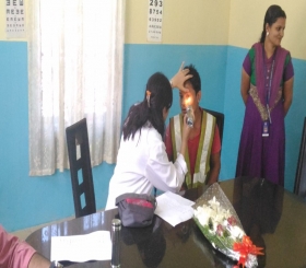
When a light flashes or a pattern moves or suddenly appears, an electrical response can be detected from the vision part of the brain. This is the visual evoked potential (VEP). The retina, which is the lining at the back of the eye, also produces an electrical signal following a flash of light. This is the electroretinogram (ERG).
Both the VEP and ERG are picked up by electrodes. These are placed on the back of the head for the VEP and on the cheek just under each eye for the ERG.
The tests can identify if a part of the visual pathway is not working properly – for example, the retina, optic nerve or brain. They can also give an estimate about the quality of pattern vision. The tests do not tell us about the interpretation of the visual information after it arrives in the brain
The VEP and ERG are especially useful in young infants or children who are unable to tell us or show us how much they can see. They are also very helpful if the doctor cannot find an obvious reason why a child sees poorly.
The tests are carried out routinely in the clinic and there are no known associated risks.

Our Mission
Establish an eye care centre with all sub-specialties in Ophthalmology. Provide clinical expertise comparable to the best in the world.